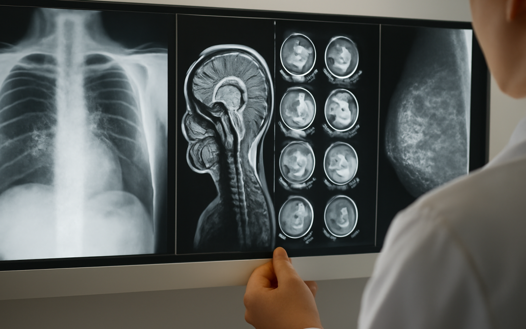
Medical imaging tests have revolutionized modern medicine by allowing healthcare professionals to peer inside the human body without making a single incision. These diagnostic tools provide critical insights into the structure and function of organs, tissues, and systems, helping to detect diseases, monitor treatment progress, and guide interventions. Understanding what medical imaging tests show is essential for both patients and practitioners to appreciate their diagnostic value and limitations. From identifying fractures to visualizing tumors and blood flow, different imaging modalities reveal unique aspects of anatomy and pathology.
One of the most common medical imaging tests is the X-ray, which utilizes ionizing radiation to create images of dense structures, particularly bones. X-rays are especially useful in evaluating fractures, dislocations, and bone infections. They can also detect lung conditions like pneumonia or chronic obstructive pulmonary disease. While X-rays offer a quick and relatively low-cost assessment of skeletal abnormalities, their utility is limited in soft tissue evaluation because many tissues have similar radiographic densities and appear indistinct. Nevertheless, this modality often serves as the first step in diagnosing injuries and conditions affecting the chest and abdomen.
Computed tomography (CT) scans provide a more detailed, cross-sectional view of the body by using X-rays taken at multiple angles and creating three-dimensional images. CT scans excel at showing both bone and soft tissues with much greater clarity than standard X-rays. They can identify tumors, internal bleeding, infections, and vascular diseases such as aneurysms or blood clots. Due to the higher radiation dose compared to conventional X-rays, CT scans are typically reserved for situations requiring intricate visualization, like trauma cases or cancer staging. Additionally, contrast agents injected into the bloodstream can further enhance the visibility of blood vessels and organs, offering valuable information about blood flow and tissue perfusion.
Magnetic resonance imaging (MRI) employs powerful magnets and radio waves to generate images of organs and tissues without exposing patients to ionizing radiation. This modality is particularly effective in displaying soft tissues such as the brain, spinal cord, muscles, and ligaments. MRI excels in diagnosing neurological disorders including multiple sclerosis, brain tumors, and stroke, as well as musculoskeletal injuries like torn ligaments and cartilage degeneration. The ability to obtain images in multiple planes and with varying tissue contrasts makes MRI indispensable for detailed assessments of complex anatomical regions. Moreover, specialized MRI techniques such as functional MRI (fMRI) can illustrate brain activity by detecting changes in blood flow, providing insight into neural function.
Ultrasound imaging uses high-frequency sound waves to create real-time images of soft tissues and organs, utilizing a device called a transducer that emits and receives sound. It is widely used to examine the abdominal organs, heart, blood vessels, and reproductive structures. One of the major advantages of ultrasound is that it involves no radiation exposure and can be performed at the bedside or in outpatient settings. For pregnant women, ultrasound offers a safe and non-invasive method to monitor fetal development and detect congenital anomalies. Additionally, Doppler ultrasound assesses blood flow in arteries and veins, helping to identify blockages or blood clots, which is crucial for managing cardiovascular diseases.
Nuclear medicine scans, such as positron emission tomography (PET) and single-photon emission computed tomography (SPECT), introduce small amounts of radioactive tracers into the body to visualize metabolic processes and physiological functions. Unlike conventional imaging that primarily shows structural information, these scans highlight areas of increased or decreased cellular activity, which is invaluable in oncology for detecting cancer spread or recurrence. PET scans are also used in cardiology to assess myocardial perfusion and viability, as well as in neurology to investigate disorders like Alzheimer’s disease by observing brain metabolism. The fusion of PET with CT or MRI further enhances diagnostic accuracy by combining functional and anatomical data into a single image.
Medical imaging tests also play an essential role in guiding minimally invasive procedures. Techniques such as fluoroscopy provide real-time X-ray imaging used during catheter placements, angiography, or joint injections. This dynamic visualization helps clinicians navigate instruments precisely, reducing risks and improving outcomes. In addition, endoscopic ultrasound combines endoscopy and ultrasound to offer detailed images of the gastrointestinal tract and surrounding tissues, aiding in the diagnosis and staging of cancers such as pancreatic or esophageal carcinoma.
Despite the impressive capabilities of these modalities, each test has inherent limitations. For example, X-rays and CT scans involve exposure to ionizing radiation, which, although minimized, can increase cancer risk if used excessively. MRI, while free from radiation, cannot be performed on patients with certain metal implants or in cases of severe claustrophobia. Ultrasound images can be limited by patient body habitus, gas interference, or operator experience. Furthermore, some abnormalities detected on imaging may be incidental findings of uncertain significance, necessitating correlation with clinical history and further tests to avoid overdiagnosis or unnecessary interventions.
Interpreting what medical imaging tests show requires specialized knowledge. Radiologists, who are expert physicians trained in reading and understanding these images, provide detailed reports that help guide clinical decisions. Their expertise is critical in differentiating normal structures from pathological changes, determining the extent and severity of disease, and recommending follow-up or additional imaging. Advances in artificial intelligence and machine learning are beginning to assist radiologists by automating detection of certain abnormalities, streamlining workflows, and reducing diagnostic errors, though human interpretation remains essential.
The growing diversity and sophistication of medical imaging tests have dramatically enhanced the precision of diagnosis and treatment planning. Conditions that once could only be inferred from symptoms and physical examination can now be visualized directly, enabling earlier detection and targeted interventions. For chronic diseases like cancer, cardiovascular disease, and neurological disorders, imaging allows for monitoring therapeutic responses and detecting complications promptly. In trauma care, rapid imaging can be lifesaving by identifying internal injuries that require surgical intervention. Moreover, imaging is fundamental in preventive medicine, facilitating screening programs such as mammography for breast cancer and low-dose CT for lung cancer in high-risk individuals.
Understanding the range of information these tests provide fosters better communication between patients and healthcare providers. Patients informed about the purpose and scope of imaging studies can engage more actively in their care, ask pertinent questions, and make informed decisions about their treatment options. Awareness of potential risks and limitations also helps set realistic expectations and alleviates anxiety related to the imaging process itself. Additionally, knowledge about how imaging contributes to diagnosis and management may encourage compliance with recommended examinations, ultimately improving health outcomes.
In recent years, innovations continue to expand what medical imaging tests can show. Hybrid imaging modalities combine functional and structural data, providing comprehensive perspectives on disease processes. Three-dimensional reconstruction and virtual reality applications offer enhanced visualization for surgical planning and education. Molecular imaging techniques aim to detect diseases at earlier molecular levels before anatomical changes occur. Continual improvements in image resolution, speed, and safety are making these diagnostic tools more accessible and valuable across various medical fields and settings.
The intricate details revealed by medical imaging tests have become indispensable for modern clinical practice. They provide a window into the anatomy and physiology of the human body, helping detect abnormalities that may not manifest clinically for some time. This capability underscores the critical role of imaging in early diagnosis, personalized therapy, and improved prognosis. As technology advances, the diagnostic scope of these tests continues to widen, promising new opportunities to unravel the complexities of human health and disease. By harnessing the full potential of medical imaging, the future of medicine looks poised to achieve more precise, safer, and more effective patient care.