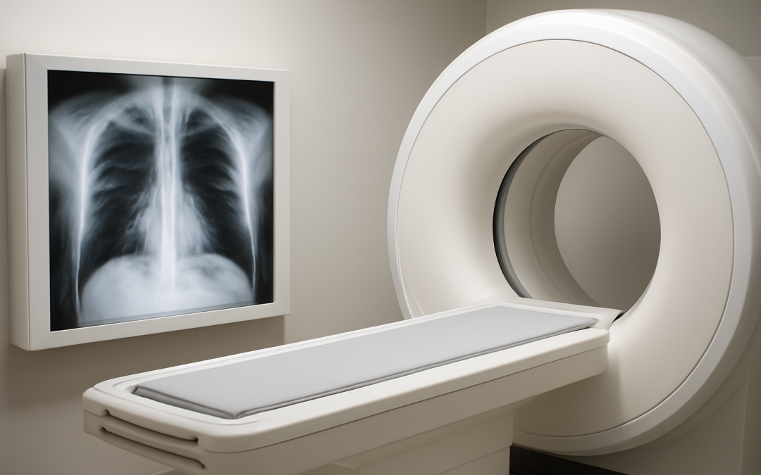
Medical imaging has revolutionized the ability to diagnose and treat a myriad of health conditions, offering insights into the human body’s internal structures without invasive procedures. Among the most commonly used imaging technologies are X-rays and magnetic resonance imaging (MRI). Both tools provide crucial diagnostic information, yet they differ fundamentally in how they work, what they reveal, and their overall application in medicine. Understanding the distinctions between X-rays and MRIs can help patients and healthcare professionals make informed decisions about which imaging technique is best suited for specific medical needs.
X-rays have been a mainstay of diagnostic imaging for over a century. They rely on electromagnetic radiation to capture images of the body’s internal structure by passing a controlled amount of radiation through the patient. Tissues that are dense, such as bones, absorb more X-rays and thus appear white or light on the resulting image, while less dense tissues like muscles or organs allow more X-rays to pass through, appearing in darker shades. This makes X-rays particularly effective for visualizing bone fractures, dental problems, and chest conditions like pneumonia or lung tumors. The speed and accessibility of X-ray imaging have made it invaluable in emergency rooms and clinics worldwide.
In contrast, MRIs expose the body to powerful magnetic fields and radio waves to generate detailed images. Unlike X-rays, MRIs do not use ionizing radiation, which makes them safer for repeated use over time, especially in vulnerable populations such as children or pregnant women. The MRI machine aligns the magnetic spin of hydrogen atoms in the body, and when external radio frequency pulses disrupt this alignment, the atoms release signals that are translated into highly detailed cross-sectional images. This method excels at rendering soft tissues with remarkable clarity, allowing for the visualization of muscles, ligaments, nervous tissue, and even the brain’s intricate structure. The detailed contrast between different types of soft tissues offered by MRI is impossible to achieve with standard X-rays.
When considering the best imaging option, it is crucial to understand the area of the body being examined. For example, X-rays are typically the first line of investigation for bone injuries, such as fractures or dislocations. They are quick, relatively inexpensive, and widely available, making them a practical choice for initial trauma assessments. Additionally, chest X-rays are often used to screen for respiratory conditions or evaluate the placement of medical devices such as catheters or pacemakers. However, the detail provided by X-rays becomes limited when assessing complex soft tissue injuries, where MRIs provide a distinct advantage.
For conditions involving soft tissues like muscles, cartilage, ligaments, tendons, or even brain anomalies, MRI is the preferred modality. This imaging technique can detect subtle changes related to inflammation, tears, or tumors that might not appear on an X-ray. In cases of neurological disorders, such as multiple sclerosis or brain tumors, MRI offers invaluable information necessary for accurate diagnosis and treatment planning. The ability to generate images in multiple planes without repositioning the patient also enhances its clinical utility. While MRI scans take longer and are generally more expensive than X-rays, their superior resolution can be critical in complex diagnostic scenarios.
The process and patient experience of undergoing an X-ray versus an MRI also differ significantly. X-ray examinations are typically brief and straightforward, often completed in just a few minutes. Patients usually stand or lie still while a single or series of images are captured, and there is minimal discomfort involved. By comparison, an MRI scan requires the patient to remain motionless inside a large, cylindrical machine for a much longer period, often between 20 to 60 minutes. The MRI environment can be noisy and confining, which may cause discomfort or anxiety for some patients, especially those with claustrophobia.
Safety considerations further distinguish X-rays and MRIs. While X-ray imaging does use ionizing radiation, the exposure level during a standard diagnostic exam is generally low and considered safe, especially when used judiciously. Nonetheless, unnecessary or repeated imaging should be avoided to minimize cumulative radiation exposure. Pregnant women are typically advised to avoid X-rays unless absolutely necessary, due to potential risks to the developing fetus. MRI, on the other hand, is free from radiation exposure, making it a safer alternative in most scenarios. However, because MRI uses strong magnetic fields, it cannot be performed on patients with certain implanted medical devices like some pacemakers or metallic implants, which could pose serious risks.
The effectiveness and accuracy of both imaging methods depend on the specific clinical question being addressed. For example, X-rays are not well suited for detecting soft tissue abnormalities such as a torn ligament or a small tumor inside an organ. In these cases, an MRI provides much more diagnostic information. Conversely, MRI is not as effective at imaging bony structures in the detail that X-rays provide. For complex fractures or bone alignment issues, X-rays remain the preferred tool. In some cases, doctors will order both an X-ray and an MRI to gain comprehensive information about an injury or disease.
In terms of cost and availability, X-rays generally represent a lower-cost option compared to MRI scans. This affordability and ease of access contribute to their frequent use in primary care and emergency settings. MRI machines are more expensive to maintain and operate, often located in specialized imaging centers or hospitals rather than smaller clinics. This difference in infrastructure can affect patient access to MRI, potentially delaying diagnosis and treatment. However, advances in technology continue to make both imaging modalities more efficient and accessible.
Interpretation of images from either modality requires expertise from radiologists who are trained to recognize subtle signs of pathology. Radiologists use X-ray and MRI findings to guide clinicians in establishing diagnoses and recommending treatment paths. While the raw images provide foundational information, radiologists’ experience is critical to discerning the clinical significance of findings and integrating imaging results with clinical symptoms and other diagnostic tests.
Technological innovations are constantly expanding the capabilities of both X-ray and MRI imaging. Digital X-rays now enhance image quality and reduce radiation dose compared to traditional film-based methods. Similarly, new MRI techniques, such as functional MRI (fMRI) and diffusion tensor imaging (DTI), provide dynamic insights into brain function and connectivity beyond static anatomical images. These advancements redefine the scope of diagnostic imaging and broaden the range of diseases and injuries that can be assessed non-invasively.
In summary, both X-rays and MRIs are powerful diagnostic tools, each with distinct advantages and limitations. X-rays excel in quickly imaging bone structures and certain chest conditions with minimal discomfort and cost, while MRIs provide detailed visualization of soft tissues without radiation exposure. Choosing between these procedures requires careful consideration of the clinical situation, patient factors, and the diagnostic information needed. Ultimately, these complementary technologies continue to enhance medical care by offering detailed, non-invasive insights that inform effective treatment and improve patient outcomes.